|
Magnetic Resonance Imaging: Basic Principles |
|||
|
|||
| . | |||
|
Case #8
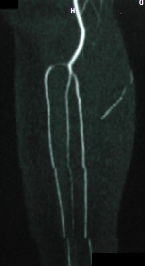
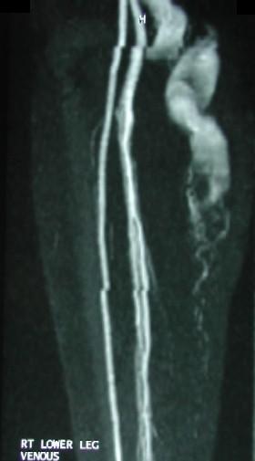
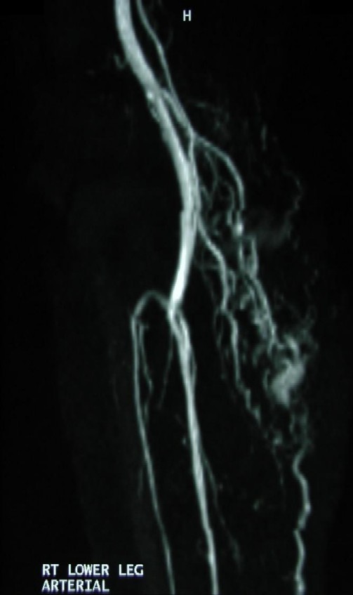
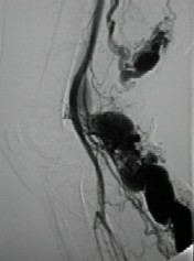
If a post-contrast MRA is performed (instead of TOF or in addition to TOF), AVM niduses can be assessed in more detail. The first two images (see above) are first and second phase post-contrast MRA images. The opacification of large draining veins is more prominent on the 2nd image. If a further delayed phase imaging was obtained, dilated veins could be more opacified, therefore, better assessed. This is clearly seen on the conventional arteriography (last image).
|
|||
|
Disclaimer: This page is intended to be an alternative source for medical professionals who deals with magnetic resonance, in clinical practice or research, but should not replace a formal education, teaching or training in the field. The author of this web source, Orhan Konez, MD, holds no responsibility for accuracy of this information, data or images. This web page does not establish any form of consultation with Dr. Konez. Information, statement or images can not be copied, duplicated and distributed in any form without permission. |
|||
|
Links: http://www.konez.com/ufe.htm - Fibroid embolization http://www.konez.com/laser.htm - Laser ablation of varicose veins http://www.konez.com/centralcatheters.htm - Central Catheters |
|||
|
|