|
Magnetic Resonance Imaging: Basic Principles |
|||||||||
|
|||||||||
| . | |||||||||
|
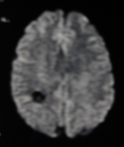
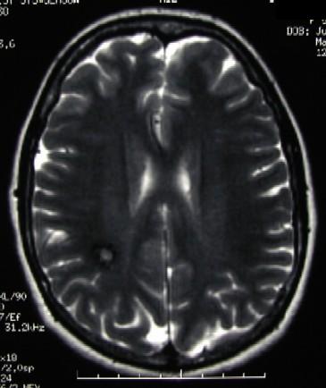
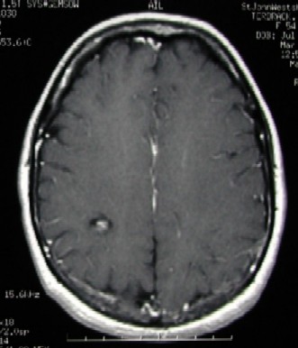
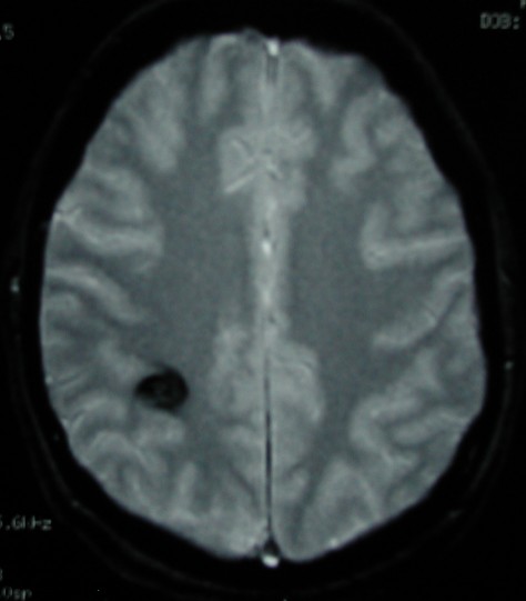
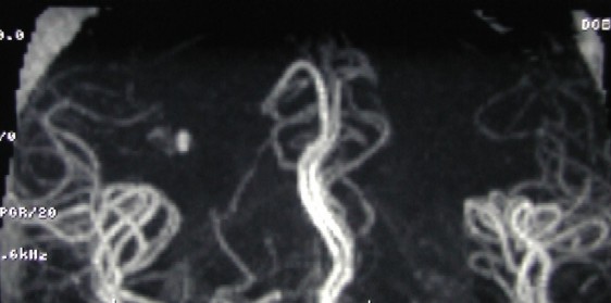
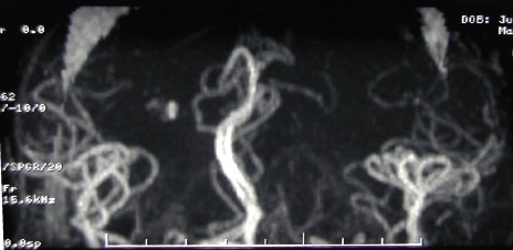
Case #7
MRI of a 47 year old patient who presented to ED with epilepsi. The first image is a diffusion weighted image and the second image is T2 weighted spin echo image (first row). The first image (second row) is a post-contrast T1 weighted image and the second image is a gradient echo image. The last two images (3rd row) are magnetic resonance angiography images obtained using the time-of-flight technique. What is your diagnosis? Findings / Diagnosis / Discussion: Diffusion weighted image shows a rounded hypointense area in the subcortical white matter, not a typical location for a hypertension related intracranial hemorrhage. This area is less noticeable on the T2 weighted spin echo image and demonstrates a mild degree contrast enhancement. No significant edema present around the lesion. The lesion is conspicuous on gradient echo image. There is a aneurysm-like signal abnormality on the TOF MR angiography images. The clinical presentation and MR findings are most consistent with a cerebral cavernous angioma. The findings are suggestive of a relatively recent bleeding into the lesion. Hypointense appearance of the lesion on the diffusion weighted image is consistent with deoxyhemoglobin status; and the aneurysm-like signal abnormality on the TOF MRA images is consistent with methamoglobin. This case is also helpful to point out the importance of the gradient echo sequence to demonstrate hemorrhage. Also, it is important to view the source images of TOF MRA to rule out aneurysms.
|
|||||||||
|
Disclaimer: This page is intended to be an alternative source for medical professionals who deals with magnetic resonance, in clinical practice or research, but should not replace a formal education, teaching or training in the field. The author of this web source, Orhan Konez, MD, holds no responsibility for accuracy of this information, data or images. This web page does not establish any form of consultation with Dr. Konez. Information, statement or images can not be copied, duplicated and distributed in any form without permission. |
|||||||||
|
Links: http://www.konez.com/ufe.htm - Fibroid embolization http://www.konez.com/laser.htm - Laser ablation of varicose veins http://www.konez.com/centralcatheters.htm - Central Catheters |
|||||||||
|
|