|
Magnetic Resonance Imaging: Basic Principles |
|||
|
|||
| . | |||
|
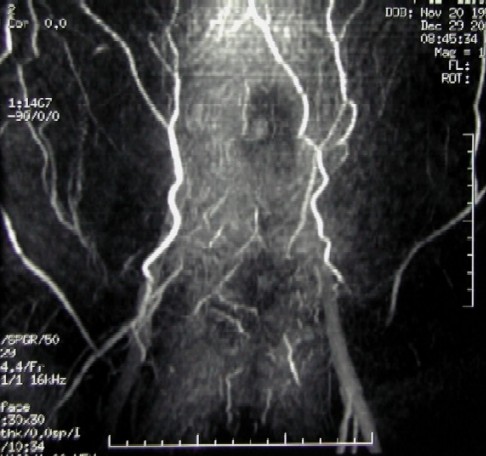
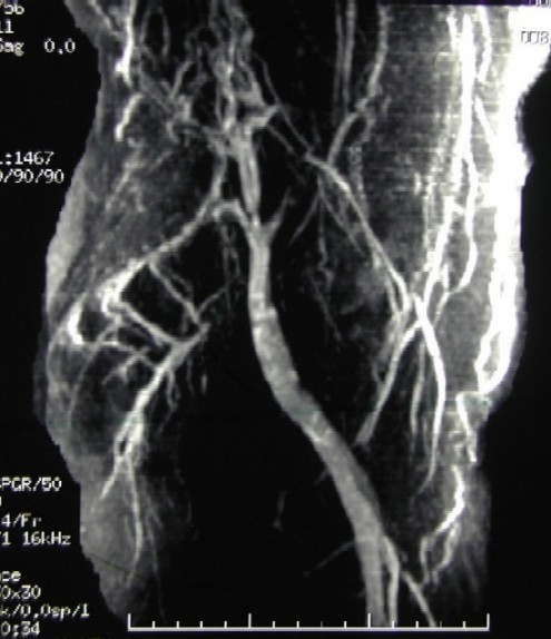
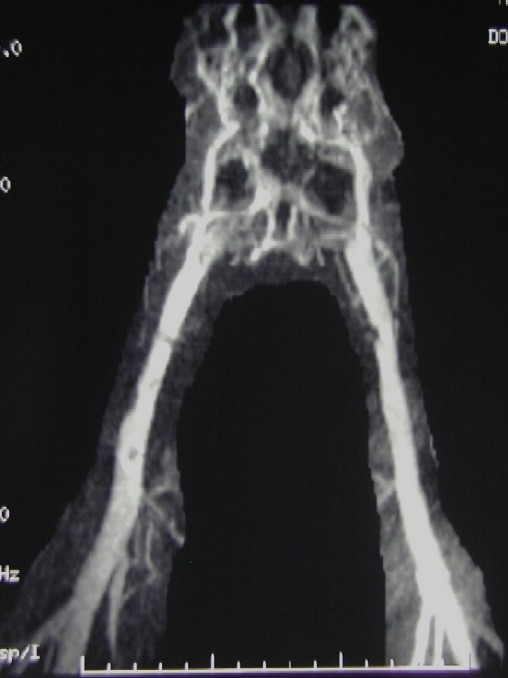
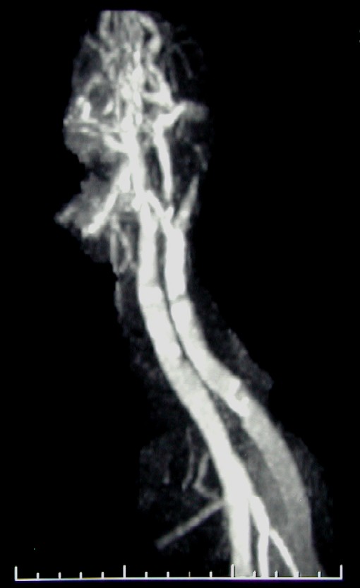
Case #5
|
|||
|
Disclaimer: This page is intended to be an alternative source for medical professionals who deals with magnetic resonance, in clinical practice or research, but should not replace a formal education, teaching or training in the field. The author of this web source, Orhan Konez, MD, holds no responsibility for accuracy of this information, data or images. This web page does not establish any form of consultation with Dr. Konez. Information, statement or images can not be copied, duplicated and distributed in any form without permission. |
|||
|
Links: http://www.konez.com/ufe.htm - Fibroid embolization http://www.konez.com/laser.htm - Laser ablation of varicose veins http://www.konez.com/centralcatheters.htm - Central Catheters |
|||
|
|