|
MRI
and MRA protocol for Vascular Anomalies
| Vascular
Anomalies:
Infantile
Hemangioma, Congenital
Hemangioma, Intramuscular
Hemangioma, Non-Involuting Hemangioma, Venous
Malformation,
Lymphatic Malformation, Lymphatic Venous
Malformation,
Arteriovenous Malformation (AVM), Arteriovenous
Fistula (AVF),
Klippel-Trenanunay
Syndrome, Parks
Weber Syndrome, Proteus Syndrome , Maffucci
Syndrome
Abbreviations:
FSE: Fast Spin-echo, PD: Proton Density, GE:
Gradient-echo, FS: Fat Suppression,
SO:
Slice Orientation, ST: Slice Thickness (mm), IT:
Imaging Time
|
|
|
|
SO |
TR/TE |
FS |
ST |
IT |
Why |
| 1 |
Axial;
Coronal and/or sagittal (+/-) |
T1
SE |
- |
3-5 |
- |
Soft
tissues (muscles, bones etc.) are assessed; to assess
if there is fat within the lesion, any hemorrhage or
blood products in or around the lesion or any flow
voids that may be related to high flow anomalies
(e.g., AVM, fistula). Also, phleboliths can be
searched within the diseased area (if phleboliths are
seen, it would be very suggestive of a venous
malformation). |
| 2 |
Axial:
Coronal and/or sagittal (+/-) |
T2-FSE
veya STIR |
+ |
3-5 |
- |
The
extent of the lesion (particularly low flow anomalies
such as venous and lymphatic malformations) and
relationship of the lesion with other soft tissue
structures can be best appreciated using this
sequence. If the lesion is a high flow anomaly (AVM,
AVF), flow voids are seen. |
| 3 |
Axial,
Coronal or sagittal |
GE,
3800/85, FA:-
No saturation
"slab"
|
- |
3-5 |
- |
Primarily
used to determine if the lesion is high- or low-flow
anomaly. If high signals are seen, the lesion is
considered to be a high flow anomaly (AVM, AVF). |
| 4 |
Axial,
Coronal or sagittal |
Post-Contrast-
MRA, 3000/120 |
- |
3-5 |
- |
In
general, this is used in addition to MRI (If not
desired, this can be skipped). It is particularly
helpful to assess the vascular architecture including
arteries and veins of the malformation. It may be
quite useful if embolization is a potential treatment
method for the lesion (AVM, fistula). |
| 5 |
Axial,
Coronal or sagittal |
Post-Contrast
T1 SE |
+ |
- |
- |
This
sequence is particularly useful to distinguish venous
malformations from lymphatic
malformations. If the
lesion shows
contrast enhancement it is considered to be
"venous", if not, it is considered a
lymphatic lesion. |
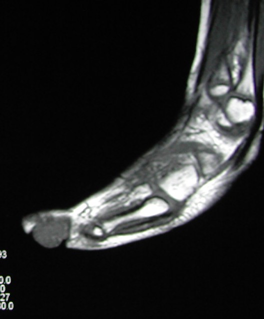
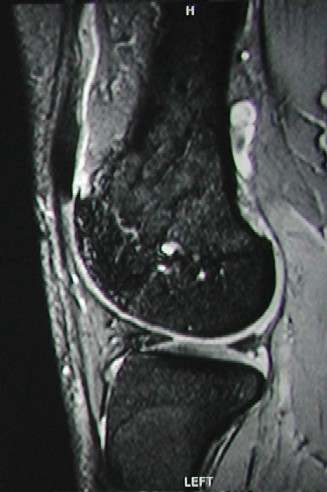
Axial
T1-SE MR images:
1st
image shows a venous malformation in the toe; the lesion can
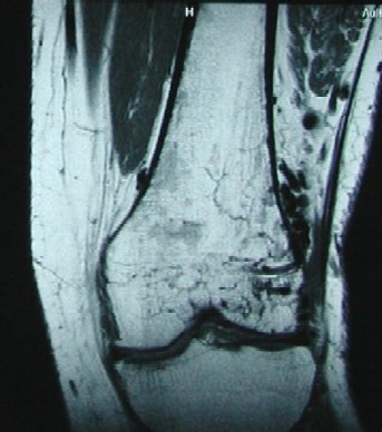
not be separated easily from other soft tissues. 2nd image
is an AVM lesion (knee). There are multiple small
hipointensities representing flow voids related to fast flow
vessels of the malformation.
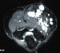
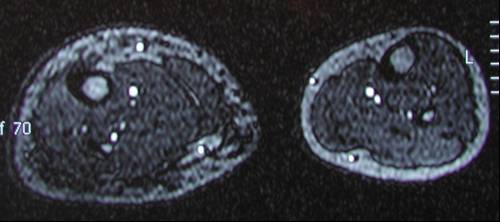
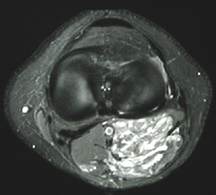
Axial
T2-FSE MR images
(with fat suppression):
1st image
shows a venous malformation in the elbow. The borders of the
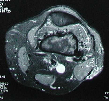
lesion can easily be determined using this sequence. 2nd
image shows an AVM in the knee, demonstrating multiple
various size signal voids in the muscles, as well as within
the bone.
Axial
GE MR images:
1st
image is a lymphatic malformation; demonstrating no signals
within the lesion. The bright signals are related to normal
extremity vessels. 2nd image shows an AVM, demonstrating
high signals due to high flow vessels.
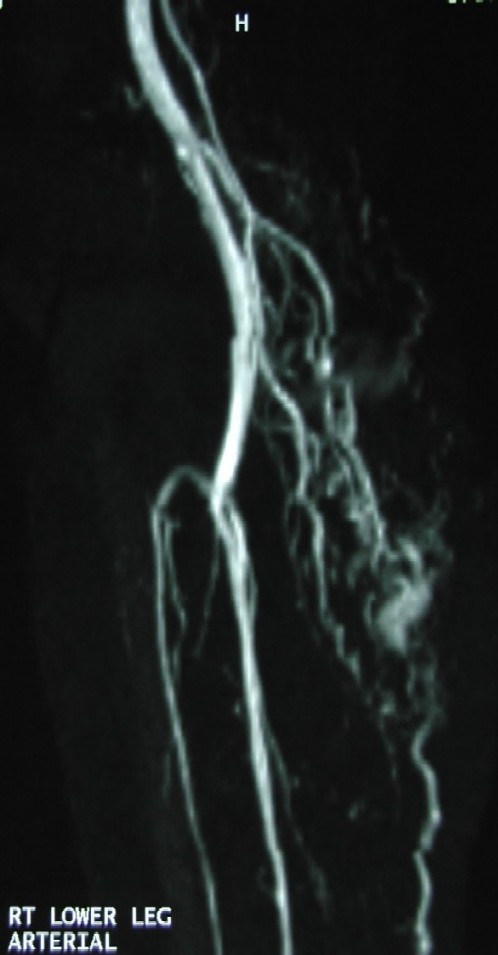
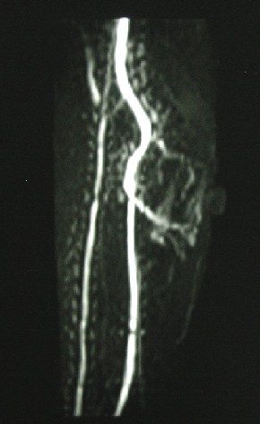
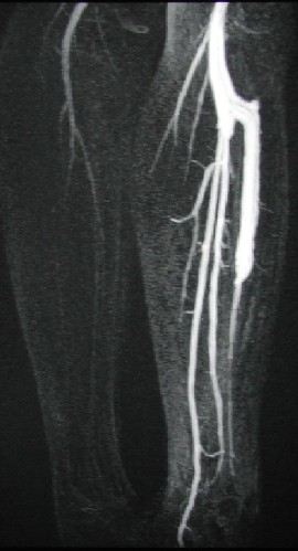
Post-Contrast
MRA
(Magnetic Resonance Angiography) images: All
images (below) are demonstrating high flow anomalies. 1st
image is an AVM in the leg, 2nd image is a lesion considered
to be intramuscular hemangioma demonstrating multiple
arterial feeders of the lesion. 3rd image demonstrates a
congenital arteriovenous fistula (AVF) in the
calf.
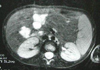
Post-contrast
T1 SE (with fat suppression)
MR images: İ
1st image is an axial
post-contrast T1 image of the knee. There is significant
contrast enhancement seen within the lesion (Venous
Malformation). Lymphatic malformations do not show contrast
enhancement or show very minimal peripheral contrast
enhancement. 2nd image shows multiple venous malformations
in the liver (Blue Rubber Bleb Nevus
Syndrome).
|