|
What
is a breast biopsy?
A breast
biopsy involves the removal of tissue from the suspicious area
of your breast. The tissue is removed by a surgeon (excisional
biopsy) or Interventional
Radiologist (Image-Guided Needle Biopsy) and sent to a
laboratory for analysis by a pathologist.
80-90% of breast biopsy results come back 'benign',
which means no cancer was detected. Still, it is essential
that you have this biopsy - it's the only way to remove the uncertainty
and get an accurate diagnosis.
What
is an Intact Breast Biopsy?
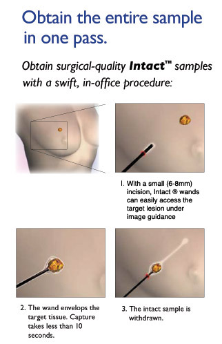
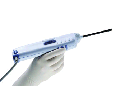
This
procedure is generally performed under local anesthesia,
causing minimal trauma to the patient's breast. It is a quick procedure, done under ultrasound or sterotactic guidance,
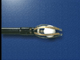
where a small device gently glides through the breast tissue
to the suspicious area or lesion. The suspicious area or
lesion is surrounded by a "capture basket", and the
suspicious tissue is removed in one pass. The tissue is then
sent to the laboratory for a diagnosis by a pathologist.
Video
Clips
|
|
 |
|
|
Prior to
the biopsy
-
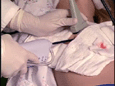
The
procedure is performed as an outpatient procedure
-
Please
wear loose-fitting clothing on the day of the procedure
-
Consult
with your physician about any medicines (e.g., Coumadin, Aspirin,
Plavix) you are taking which can prevent clotting
What to
expect during the biopsy
-
The
longest portion of the procedure is the preparation. The actual
biopsy - removal of
the suspicious tissue or lesion - takes less than 10 seconds.
-
You will
have a cold pad on your back, which is necessary for the Intact
system to function
-
During
the procedure, you may feel pressure, tingling, or a 'pinch' in
your breast. Plenty of anesthetic is used to keep the you comfortable
-
You
may also notice an unusual smell during the the biopsy. This smell
is a normal part of the procedure
-
You will
hear various sounds during the procedure. The machine will beep
and you will hear something that sounds like a hair dryer while
the tissue is being removed
-
A tiny
marker (visible under X-ray and Ultrasound) is placed into the
biopsy site at the end of the procedure
What to
expect after the procedure
-
The small incision is generally covered with a steri-strip bandage
-
After the procedure is completed, you will be given
post-biopsy care instructions
-
You may experience some slight swelling, bruising and
discharge, which is normal. Any discomfort should disappear in 1-2
days. If it persists, call your doctor
-
Consult
with your doctor if you have a fever, or significant bleeding, or
redness at the biopsy site
-
The
report from the pathologist will be sent to your doctor in a few
days. Your doctor will call to schedule time to discuss the
results with you
Advantages
of the Intact BLES technique:
Significantly
improved sampling accuracy: The needle is easily visible under real-time
ultrasound and the system captures
significantly larger size tissue than typical core or suction
biopsy needles; therefore, false negative results should be
significantly less.
Less
complications: Because
of the Radio Frequency technology used to advance the needle and also the capture
the suspicious tissue, bleeding is less of a concern than that of
typical needle biopsies. Also, capturing the suspicious lesion with
the BLES technique is a slow process and can be seen (real time) under
ultrasound; because there is no fast
motion of the needle tip complications such as needle
penetration into the chest wall or other normal tissues are a less
likely possibility.
A marker at the biopsy site
can be placed more reliably: The BLES
system creates a small cavity at the biopsy site; therefore,
placement of a marker is a much easier process
and more importantly can be done more accurately.
Potential
theoretical risk of disturbing the potential cancer tissue is
unlikely:
Since the BLES technique captures the tissue rather than directly
penetrating the tissue, the potential cancer tissue (at least large portion
of it) is removed without much
disturbance in the area.
|