|
Magnetic Resonance Imaging: Basic Principles |
|
|||||||||
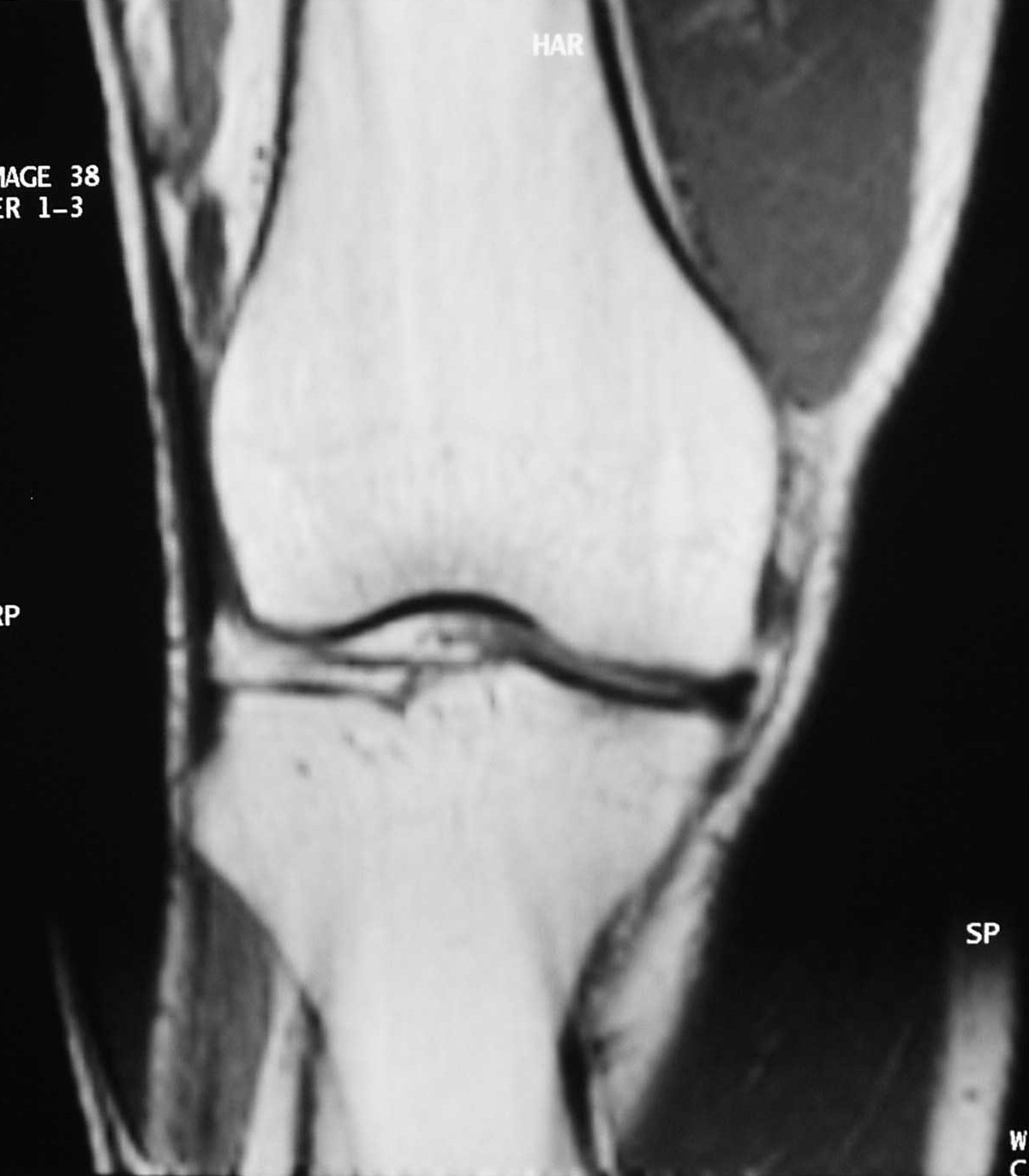
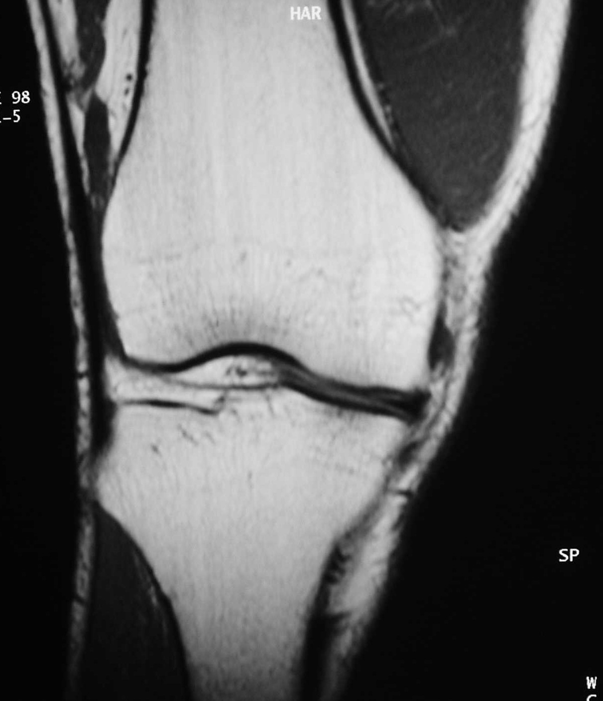
Findings / Diagnosis / Discussion: There are bilateral soft tissue signal abnormalities seen in the lower section of the image. This artifact is known as "Wrap Around artifact" or "Aliasing". The cause of this artifact is to use a small field of view (FOV), resulting in undersampling in k-space.
|
|||||||||
| Disclaimer: This page is intended to be an alternative source for medical professionals who deals with magnetic resonance, in clinical practice or research, but should not replace a formal education, teaching or training in the field. The author of this web source, Orhan Konez, MD, holds no responsibility for accuracy of this information, data or images. This web page does not establish any form of consultation with Dr. Konez. Information, statement or images can not be copied, duplicated and distributed in any form without permission. | |||||||||
|
|