|
Magnetic Resonance Imaging: Basic Principles |
|
||||||
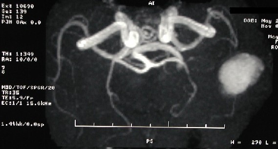
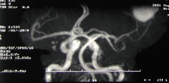
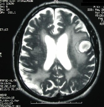
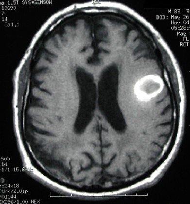
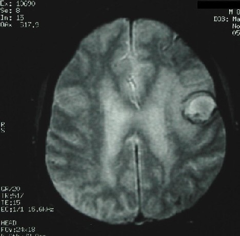
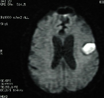
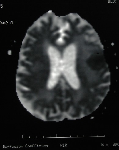
Diagnosis: TOF-MRA (arteriography) artifact (due to a relatively acute intracranial hemorrhage) simulating a large intracranial aneurysm. Discussion: 1- Acute-subacute hemorrhage can cause an MRA artifact, and if not carefully assessed, it may be misdiagnosed as an aneurysm. Methemoglobin within thrombus has a short T1 relaxation time and cause high signal on TOF images, simulating intraluminal flow. Phase-contrast angiography technique is not effected by methemoglobin and can be used to distinguish thrombus from flow. However, both TOF and phase-contrast techniques are subject to signal loss from the magnetic susceptibility effects of deoxyhemoglobin and hemosiderin/ferritin. This is mostly a problem in partially thrombosed aneursysms, patent lumen of the aneurysm may be obscured. This is not a commonly encountered MRA artifact. Meticulous review of the whole MRI exam and review of the clinical history are important. Intensity of the intracranial blood changes allowing to estimate the age of the hematoma. Based on the above seen signal characteristics, this patient has a subacute intracerebral hematoma. 2- Intracerebral hematoma can be seen as an hyperintense area on diffusion weighted imaging. Therefore, diffusion weighted imaging does not have a significant diagnostic value in this clinical setting. However, the technique may be useful to investigate possible ischemic areas. The following lesions can be hyperintens on diffusion weighted imaging: acute stroke, epidermoid, pyogenic infection, herpes encephalitis, Creutzfeldt-Jacob syndrome, diffuse axonal injury, oxyhemoglobin, extracellular methemoglobin and some MS lesions.
|
||||||
|
|
||||||
| Disclaimer: This page is intended to be an alternative source for medical professionals who deal with magnetic resonance, in clinical practice or research, but should not replace a formal education, teaching or training in the field. The author of this web source, Orhan Konez, MD, holds no responsibility for the accuracy of this information, data or images. This web page does not establish any form of consultation with Dr. Konez. Information, statements or images can not be copied, duplicated or distributed in any form without permission. | ||||||
|
|